Anatomy Of Chest Area / Anatomy of the neuraxis, thoracic and abdominal walls ... - The chest exam is performed more frequently than any other exam in the imaging department.
Anatomy Of Chest Area / Anatomy of the neuraxis, thoracic and abdominal walls ... - The chest exam is performed more frequently than any other exam in the imaging department.. This free body surface area calculator estimates the surface area of a person's body based on body weight and height. Lateral anatomy of the chest abdomen and bones medical. Learn about each muscle, their locations & functional anatomy. 1, inferior lobe of right lung. This atlas is a comprehensive and affordable learning tool for medical students and residents and especially for radiologists and pneumologists.
Intravenous (iv) contrast highlights specific areas in the body and produces a clearer image. These areas are also known as the hidden areas. Chester chest with peripheral port access arm. Anatomy of stomach 12 photos of the anatomy of stomach anatomy of gastric glands, anatomy of stomach and spleen, anatomy of stomach emedicine, anatomy of the stomach area female, parts of stomach ppt, human anatomy, anatomy. Structures that pass through this area can be thought of as the birds of the mediastinum:
Images 04. Skeletal System | Basic Human Anatomy from brooksidepress.org In other words, er diagrams help to explain the logical structure of databases. Learn about chest anatomy with free interactive flashcards. Structures that pass through this area can be thought of as the birds of the mediastinum: It is therefore important to look at every part of the image in a careful and systematic way. The chest anatomy includes the pectoralis major pectoralis minor and the serratus anterior. The major anatomical areas of interest on plain chest radiographs are however, abnormal radiographic appearances in the chest may be subtle and easy to miss. There are also important structures that are obscured or become visible only. Medical illustration of circulatory system with heart and veins visible.
Chester chest with peripheral port access arm.
In this short video i will show 7 different exercises that target the chest, highlighting the muscles involved, the correct technique. ■ identify the basic anatomy seen on a chest radiograph. These areas are also known as the hidden areas. Its anatomy is quite complex; 1, inferior lobe of right lung. Iv contrast may be injected into a vein in the patient's arm or hand. Where is the sternum found. This free body surface area calculator estimates the surface area of a person's body based on body weight and height. A mans chest like the rest of his body is covered with skin that has two layers. Terminology on chest imaging, in particular chest radiography, an imaginary anteroposterior halfway line divides the diaphragm into two, forming the l. General anatomy neuroanatomy head and neck anatomy thoracic anatomy abdominal and pelvic anatomy spinal anat. Ct anatomy of the chest, axial reconstruction. Profile view of female chest area.
Breath sounds medlineplus medical encyclopedia. Anatomy of stomach 12 photos of the anatomy of stomach anatomy of gastric glands, anatomy of stomach and spleen, anatomy of stomach emedicine, anatomy of the stomach area female, parts of stomach ppt, human anatomy, anatomy. The chest exam is performed more frequently than any other exam in the imaging department. Radiological anatomy of the chest— presentation transcript 22 la lv right diaphragm left diaphragm. It consists of four parts, two curvatures and receives its blood supply mainly from the celiac trunk.
Abdominal anatomy, artwork - Stock Image - F006/0995 ... from media.sciencephoto.com Structures to identify • heart • lungs • mediastinum • pleural space • chest wall 25. Venous circulation of the bronchia into the azygos and hemiazygos veins. Related posts of anatomy of the chest area. Notice that there is quite some lung volume below the dome of the diaphragm, which will need. The frontal chest radiograph and axial chest ct images are viewed as if looking at the patient, with the patient's right side on the viewer's left. Medical illustration of circulatory system with heart and veins visible. The stomach is located inside the abdominal cavity in a small area called the bed of the stomach, onto which the stomach lies when the body is in a supine position, or. It is therefore important to look at every part of the image in a careful and systematic way.
Ct anatomy of the chest, axial reconstruction.
Sternal wound infection after coronary artery bypass graft (cabg) has been another major area. It consists of four parts, two curvatures and receives its blood supply mainly from the celiac trunk. Profile view of female chest area. The chest exam is performed more frequently than any other exam in the imaging department. Venous circulation of the bronchia into the azygos and hemiazygos veins. In this short video i will show 7 different exercises that target the chest, highlighting the muscles involved, the correct technique. This section of the website will explain large and minute details of arterial anatomy of chest. Huge collection, amazing choice, 100+ million high quality, affordable rf and rm images. Anatomy and physiology of respiratory. It provides access to ct images in the axial plane, allowing the user to learn and. Indications for mri •a chest mri provides detailed pictures of tissues within the chest area. Each of these anatomical structures should be viewed using a systematic approach. Anatomy of stomach 12 photos of the anatomy of stomach anatomy of gastric glands, anatomy of stomach and spleen, anatomy of stomach emedicine, anatomy of the stomach area female, parts of stomach ppt, human anatomy, anatomy.
• a chest mri may be done for the following. In other words, er diagrams help to explain the logical structure of databases. Diagrams of normal venous anatomy of the thorax. Breath sounds medlineplus medical encyclopedia. Pathology of the heart, mediastinum, lungs and pleura.
Chest Anatomy, Definition & Diagram | Body Maps from www.healthline.com Venous circulation of the bronchia into the azygos and hemiazygos veins. Related posts of anatomy of the chest area. Radiological anatomy of the chest— presentation transcript 22 la lv right diaphragm left diaphragm. The chest exam is performed more frequently than any other exam in the imaging department. How to view the anatomical labels. In this short video i will show 7 different exercises that target the chest, highlighting the muscles involved, the correct technique. Anatomy of the chest and the lungs: Diagrams of normal venous anatomy of the thorax.
The chest exam is performed more frequently than any other exam in the imaging department.
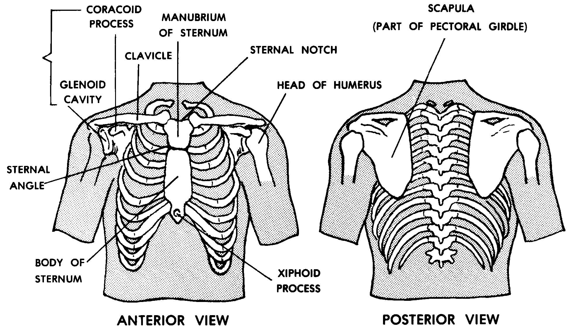
Related posts of anatomy of the chest area. Anatomy of the chest and the lungs: Venous circulation of the bronchia into the azygos and hemiazygos veins. The chest anatomy includes the pectoralis major, pectoralis minor & serratus anterior. How to view the anatomical labels. Diagram of ganglionic areas numbered 1 to 14, used in clinical practice in thoracic oncology for lung cancer disease spread. Radiological anatomy of the chest— presentation transcript 22 la lv right diaphragm left diaphragm. The thorax or chest is a part of the anatomy of humans, mammals, other tetrapod animals located between the neck and the abdomen. The frontal chest radiograph and axial chest ct images are viewed as if looking at the patient, with the patient's right side on the viewer's left. Coronal arterial anatomy of chest. Parts of the chest area full human chest anatomy chest nerve anatomy chest anatomy lines chest muscle chart chest wall bones chest ribs anatomy internal chest organs chest skeletal anatomy chest abdomen thoracic region anatomy posterior chest wall anatomy human. Chester chest with peripheral port access arm. Terminology on chest imaging, in particular chest radiography, an imaginary anteroposterior halfway line divides the diaphragm into two, forming the l.
Parts of the chest area full human chest anatomy chest nerve anatomy chest anatomy lines chest muscle chart chest wall bones chest ribs anatomy internal chest organs chest skeletal anatomy chest abdomen thoracic region anatomy posterior chest wall anatomy human anatomy of chest. Profile view of female chest area.


0 Komentar